GINGIVAL AND PALATAL CYSTS OF THE NEWBORN AND ADULT
Features in the Newborn
Gingival cysts of the newborn are often multiple sessile domeshaped lesions measuring about 2 to 3 mm in diameter. They are chalk white and present predominantly on the maxillary anterior alveolar ridge just lingual to the crest.
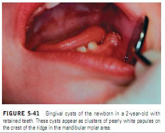
Those in the posterior region of the jaw are found directly on the crest of the ridge occlusal to the crowns of the molar teeth (Figure 5-41). These lesions are usually seen in newborn or very young infants and disappear shortly after birth; they are thought to originate from remnants of the dental lamina. These cysts tend to rupture and disappear spontaneously. The eponyms “Epstein’s pearls” and “Bohn’s nodules” have both been used to describe odontogenic cysts of dental lamina origin, but these terms are not considered to be accurate. Epstein originally described keratin-filled nodules found along the midpalatal region, probably derived from entrapped epithelium along the lines of fusion of the palatal processes. These are considered quite rare. Bohn’s nodules are thought to be keratinfilled cysts scattered across the palate but most plentiful along the junction of the hard palate and soft palate and are thought to be derived from palatal salivary glands. Bohn’s nodules probably relate to what are presently called gingival cysts of the newborn. Krisover reported finding 65 examples of gingival cysts in 17 infants.Features in the Newborn
Gingival cysts of the newborn are often multiple sessile domeshaped lesions measuring about 2 to 3 mm in diameter. They are chalk white and present predominantly on the maxillary anterior alveolar ridge just lingual to the crest.
The incidence in Japanese infants was almost 90%, an incidence considered significantly higher than that seen in black or white newborns.Palatal cysts of the newborn occasionally persist into adult life and appear as peripheral odontogenic keratocysts.
Features in the Adult
Gingival cysts of the adult are thought to arise from dental lamina rests or from entrapment of surface epithelium.
They are most common in the canine and premolar area of
Features in the Adult
Gingival cysts of the adult are thought to arise from dental lamina rests or from entrapment of surface epithelium.
They are most common in the canine and premolar area of
the mandible and maxillary lateral incisor area and usually occur during the fifth and sixth decades of life. They have a very strong resemblance to lateral periodontal cysts, and there is a strong correlation between these two types of lesions. Patients have had lateral periodontal cysts subsequent to the development of a gingival cyst,and lateral periodontal cysts are thought to be the intrabony counterpart of the gingival cyst. Gingival cysts usually appear as sessile painless growths involving the interdental area of the attached gingiva. These lesions often appear to be white or yellow white to blue and measure about 0.5 to 1 cm in diameter. They occasionally cause some superficial destruction of the underlying bone. A definite radiolucency is thought to represent a lateral periodontal cyst


