Radiography with the use of contrast agents is still performed in some facilities, but its usage has decreased significantly with the evolution of advanced imaging techniques. The major contrast-enhanced examinations used in dentistry are arthrography and sialography.
In arthrography of the TMJ, radiopaque material is injected into the lower (and sometimes also the upper) joint space under fluoroscopic guidance. Once the dye is in place, fluoroscopic recordings of the joint in motion may be made in order to assess the shape, location, and function of the articular disk.
Radiography or tomography may also be performed afterwards (Figure 3-14). Arthrography is invasive and technically difficult and has been replaced by MRI in most institutions.
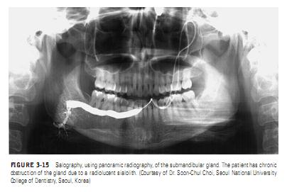
In sialography, contrast medium is injected into the major duct of the salivary gland of interest. The distribution of the ductal system, along with any patterns such as narrowing or dilation of ducts or such as contrast extravasation or retention, can provide helpful information regarding the inflammatory, obstructive, or neoplastic condition affecting the gland
(Figure 3-15). In many institutions, CT, MRI, or US is used more often than sialography to evaluate the salivary glands.

