Chewing tobacco is an important established risk factor for the development of oral carcinoma in the United States.
Habitually chewing tobacco leaves or dipping snuff results in the development of a well-recognized white mucosal lesion in the area of tobacco contact, called smokeless tobacco keratosis, snuff dipper’s keratosis, or tobacco pouch keratosis.
Habitually chewing tobacco leaves or dipping snuff results in the development of a well-recognized white mucosal lesion in the area of tobacco contact, called smokeless tobacco keratosis, snuff dipper’s keratosis, or tobacco pouch keratosis.
While these lesions are accepted as precancerous, they are significantly different from true leukoplakia and have a much lower risk of malignant transformation.This habit was once almost universal in the United States and is very common among certain other populations, most notably in Sweden, India, and Southeast Asia.
Smokeless tobacco use among white males in the United States has shown a recent resurgence.The estimated proportion of adult men in the United States who regularly use “spit” tobacco ranges from 6 to 20%.This range is attributed to significant geographic, cultural, and gender variations in chewing habits.The cumulative incidence for smokeless tobacco use was highest for non-Hispanic white males.Unfortunately, the habit starts relatively early in life, usually between the ages of 9 and 15 years, and is rarely begun after 20 years of age. Recent epidemiologic data indicate that over five million Americans use smokeless tobacco and that more than 750,000 of these users are adolescents. It is estimated that each year in the United States, approximately 800,000 young people between the ages of 11 and 19 years experiment with smokeless tobacco and that about 300,000 become regular users.Smokeless tobacco contains several known carcinogens, including N-nitrosonornicotine (NNN), and these have been proven to cause mucosal alterations.In addition to its established role as a carcinogen, chewing tobacco may be a risk factor in the development of root surface caries and, to a lesser extent, coronal caries. This may be due to its high sugar content and its association with increased amounts of gingival recession.The duration of exposure is very important in the production of mucosal damage. Leukoplakia has been reported to develop with the habitual use of as little as three cans of snuff per week for longer than 3 years.Although all forms of smokeless tobacco may result in mucosal alterations, snuff (a finely powdered tobacco) appears to be much more likely to cause such changes than is chewing tobacco.
Smokeless tobacco is not consistently associated with increased rates of oral cancer.Approximately 20% of all adult Swedish males use moist snuff, yet it has not been possible to detect any significant increase in the incidence of cancer of the oral cavity or pharynx in Sweden. By international standards, the prevalence of oral cancer is low in Sweden.This has been attributed to variations in the composition of snuff, in particular, the amount of fermented or cured tobacco in the mixture. The carcinogen NNN is present in much lower concentrations in Swedish snuff, probably because of a lack of fermentation of the tobacco. Also, the high level of snuff use might decrease the amount of cigarette smoking and therefore lead to a lesser prevalence of oral cancer.
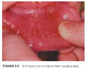
TYPICAL FEATURESNumerous alterations are found in habitual users of smokeless tobacco. Most changes associated with the use of smokeless tobacco are seen in the area contacting the tobacco. The most common area of involvement is the anterior mandibular vestibule, followed by the posterior vestibule.The surface of the mucosa
appears white and is granular or wrinkled (Figure 5-9); in some cases, a folded character may be seen (tobacco pouch keratosis). Commonly noted is a characteristic area of gingival recession with periodontal-tissue destruction in the immediate area of contact (Figure 5-10). This recession involves the facial aspect of the tooth or teeth and is related to the amount and duration of tobacco use. The mucosa appears gray or gray-white and almost translucent. Since the tobacco is not in the mouth during examination, the usually stretched mucosa appears fissured or rippled, and a “pouch” is usually present. This white tobacco pouch may become leathery or nodular in long-term heavy users (Figure 511). Rarely, an erythroplakic component may be seen. The lesion is usually asymptomatic and is discovered on routine examination. Microscopically, the epithelium is hyperkeratotic and thickened. A characteristic vacuolization or edema may be seen in the keratin layer and in the superficial epithelium. Frank dysplasia is uncommon in tobacco pouch keratosis.
TREATMENT AND PROGNOSIS
Cessation of use almost always leads to a normal mucosal appearance within 1 to 2 weeks.Biopsy specimens should be obtained from lesions that remain after 1 month. Biopsy is particularly indicated for those lesions that appear clinically atypical and that include such features as surface ulceration, erythroplakia, intense whiteness, or a verrucoid or papillary surface.The risk of malignant transformation is increased fourfold for chronic smokeless tobacco users.
Cessation of use almost always leads to a normal mucosal appearance within 1 to 2 weeks.Biopsy specimens should be obtained from lesions that remain after 1 month. Biopsy is particularly indicated for those lesions that appear clinically atypical and that include such features as surface ulceration, erythroplakia, intense whiteness, or a verrucoid or papillary surface.The risk of malignant transformation is increased fourfold for chronic smokeless tobacco users.


