Lichenoid reactions and lichen planus exhibit similar histopathologic features.Lichenoid reactions were differentiated from lichen planus on the basis of (1) their association with the administration of a drug, contact with a metal, the use of a food flavoring, or systemic disease and (2) their resolution when the drug or other factor was eliminated or
when the disease was treated.Clinically, lichenoid lesions may exhibit the classic appearance of lichen planus, but atypical presentations are seen, and some of the dermatologic lesions included in this category show little clinical lichenification.Table 5-3 lists some of the disorders that are currently proposed as lichenoid reactions.Drug-Induced Lichenoid ReactionsDrug-induced lichenoid eruptions include those lesions that are usually described in the dental literature under the topic of lichenoid reactions (ie, oral mucosal lesions that have the clinical and histopathologic characteristics of lichen planus, that are associated with the administration of a drug, and that resolve following the withdrawal of the drug).A drug history can be one of the most important aspects of the assessment of a patient with an oral or oral-and-skin
lichenoid reaction. Clinically, there is often little to distinguish drug-induced lichenoid reactions from lichen planus. However, lichenoid lesions that include the lip and lichenoid lesions that are symmetric in distribution and that also involve the skin are more likely to be drug related.
Histopathologically, lichenoid drug eruptions may show a deep as well as superficial perivascular lymphocytic infiltrate rather than the classic bandlike infiltrate of lichen planus, and eosinophils, plasma cells, and neutrophils may also be present in the infiltrate.
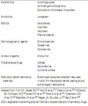
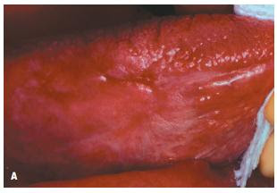
Drug-induced lichenoid reactions may resolve promptly when the offending drug is eliminated. However, many lesions take months to clear; in the case of a reaction to gold salts, 1 or 2 years may be required before complete resolution. Gold therapy, nonsteroidal anti-inflammatory drugs (NSAIDs), diuretics, other antihypertensives, and oral hypoglycemic agents of the sulfonylurea type are all important causes of lichenoid reactions (Table 5-4; Figure 5-36).
216–224
(Because more drugs are being added to it on a continual basis, this list should by no means be considered complete.)
Surveys of medication use among patients with various oral keratoses, including lichen planus, have found the use of antihypertensive drugs to be more common among lichen planus patients.
219
The use of NSAIDs was 10 times more frequent among those with erosive lichen planus.
Penicillamine is associated with many adverse reactions, including lichenoid reactions, pemphigus-like lesions, lupoid reactions and stomatitis, and altered taste and smell functions.
220,223
A number of the drugs that have been associated with lichenoid reactions may also produce lesions of discoid lupus erythematosus (lupoid reactions).
Graft-versus-Host Disease
Graft-versus-host disease (GVHD)
216
225–240
is a complex multisystem immunologic phenomenon characterized by the interaction of immunocompetent cells from one individual (the donor) to a host (the recipient) who is not only immunodeficient but who also possesses transplantation isoantigens foreign to the graft and capable of stimulating it.
Reactions of this type occur in up to 70% of patients who undergo allogenic bone marrow transplantation, usually for treatment of refractory acute leukemia. There may be both acute (< 100 days after bone marrow transplantation) and chronic (after day 100, post transplantation) forms of the condition. The pathogenesis is probably related to an antigen-dependent proliferation of transplanted donor T-cell lymphocytes that are genetically disparate from the recipient’s own tissues and that give rise to a generation of effector cells that react with and destroy recipient tissues. Of these, the epidermal (skin and mucous membrane) lesions are often most helpful clinically in establishing a diagnosis.
CLINICAL FEATURES
The epidermal lesions of acute GVHD range from a mild rash to diffuse severe sloughing. This may include toxic epidermal necrolysis (Lyell’s disease), a type of erythema multiforme in
which large flaccid bullae develop with detachment of the epidermis in large sheets, leaving a scalded skin appearance.
Oral mucosal lesions occur in only about one-third of cases and are only a minor component of this problem.
Chronic GVHD is associated with lichenoid lesions that affect both skin and mucous membranes. Oral lesions occur in
Oral mucosal lesions occur in only about one-third of cases and are only a minor component of this problem.
Chronic GVHD is associated with lichenoid lesions that affect both skin and mucous membranes. Oral lesions occur in
80% of cases of chronic GVHD; salivary and lacrimal gland epithelium may also be involved.In some cases, the intraoral lichenoid lesions are extensive and involve the cheeks, tongue, lips, and gingivae. In most patients with oral GVHD, a fine reticular network of white striae that resembles OLP is seen. Patients may often complain of a burning sensation of the oral mucosa. Xerostomia is a common complaint due to the involvement of the salivary glands. The development of a pyogenic granuloma on the tongue has been described as a component of chronic GVHD.The mouth is an early indicator of a variety of reactions and infections associated with transplantation-related complications. The majority of these infections are opportunistic Candida infections although infections with other agents (such as gram-negative anaerobic bacilli and Aspergillus) have also been described.The differential diagnosis of oral lesions that develop in patients some months after bone marrow transplantation thus includes candidiasis and other opportunistic infections, toxic reactions to chemotherapeutic drugs that are often used concomitantly, unusual viral infections (herpesvirus, Cytomegalovirus), and lichenoid drug eruptions.Because of the potential involvement of salivary glands in chronic GVHD, biopsy of minor salivary glands is a useful diagnostic procedure in some cases.The histopathologic features of chronic GVHD may resemble those of OLP.
TREATMENT AND PROGNOSIS
The principle basis for management of GVHD is prevention by careful histocompatibility matching and judicious use of immunosuppressive drugs. In some cases, topical corticosteroids and palliative medications may facilitate the healing of the ulcerations. Ultraviolet A irradiation therapy with oral psoralen has also been shown to be effective in treating resistant lesions.Topical azathioprine suspension has been used as an oral rinse and then swallowed, thereby maintaining the previously prescribed systemic dose of azathioprine. This resulted in improvement in cases of oral GVHD that was resistant to other approaches to management. Topical azathioprine may provide additional therapy in the management of immunologically-mediated oral mucosal disease.Clinicians evaluating cases of lichen planus should rule out the possibility of oral lichenoid reactions. Lichenoid drug reactions manifesting in the oral cavity have received some attention, and oral lichenoid GVHD has been well described, but it is likely that oral lichen planus as currently diagnosed also represents a heterogeneous group of lesions that require more specific identification and customized focused treatment.


