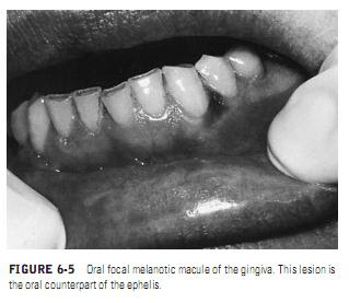
Unlike ephelides and melanotic macules, which result from an increase in melanin pigment synthesis, nevi are due to benign proliferations of melanocytes.There are two major types, based on histology, and these two types tend to show differences clinically as well, particularly in tint and
coloration. Nevocellular nevi arise from basal-layer melanocytes early in life. In the evolutionary stages, the nevus cells maintain their localization to the basal layer, residing at the junction of the epithelium and the basement membrane and underlying connective tissue. Since proliferation is minimal, these nevi are macular and are classified as junctional nevi. In general, they are flat and brown and have a regular round or oval outline. With time, the melanocytes form clusters at the epitheliomesenchymal junction and begin to proliferate down into the connective tissue although they do not invade vessels or lymphatics. Such nevi assume a domeshaped appearance (since more cells have accumulated) and are referred to as compound nevi. In late puberty, the melanocytes (now known as nevus cells) in compound nevi lose their continuity with the surface epithelium, and the cells become localized to the deeper connective tissues. They are then termed intradermal nevi when on skin and intramucosal nevi when in the mouth. On the skin, they are elevated brown nodules that often have hair protruding from them. Thus, in adults, junctional nevi should not exist. When a nevus shows microscopic evidence of junctional activity, premelanomatous change should be suspected.
The second type of nevus, not derived from basal-layer melanocytes, is the blue nevus. The blue nevus is blue on the skin because the melanocytic cells reside deep in the connective tissue and because the overlying vessels dampen the brown coloration of melanin, yielding a blue tint. The melanocytes of a blue nevus differ morphologically from those of a nevocellular nevus by being more spindle shaped while containing significant amounts of pigment. Such cells are neuroectodermally derived yet are believed to represent cells that failed to reach the epithelium. A rare cellular form of blue nevus also exists, and neither the ordinary nor the cellular form has the potential to become a melanoma.
In the oral mucosa, both nevocellular and blue nevi tend to be brown and may be macular or nodular (Figure 6-6). They may be seen at any age and are found most frequently on the palate and gingiva but may also be encountered in the buccal mucosa and on the lips. Once they reach a given size, their growth ceases, and the lesions remain static. Biopsy is necessary for diagnostic confirmation since the clinical diagnosis includes many other focal pigmentations, such as melanotic macule, melanoma, and amalgam tattoo. Simple excision is the treatment of choice.
